
Heart-related diseases are the leading cause of death in the industrialized world. Cardiac stem cell therapy is a promising new way of reducing those numbers, but its application has proven to be less effective than hoped. Now researchers at Stanford University have developed nanoparticles that can be used to image stem cells implanted into the heart. They claim this will help improve the efficiency of these transplants drastically.
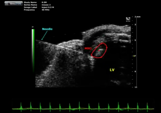
Stem cell therapy uses cells that have the ability to transform into a wide variety of mature cell types. When implanted in the heart, for instance, they can transform into heart cells. This ability can be used to repair injured or diseased parts of the heart. Sadly, current methods of introducing stem cells rely on trial and error.
Let’s say, for instance, that a patient suffers a heart attack, which leaves some of his heart cells injured. To help the heart heal, the patient is first put into an MRI scanner to locate the areas of the heart that need repair. Once those are determined, doctors use the scan to implant new stem cells into these regions. After implantation, the patient is returned to the MRI to determine the location and number of implanted cells—if they’re not where they need to be, the patient is returned to surgery. This is exhaustively repeated.
Read 6 remaining paragraphs | Comments
from Ars Technica