
Earlier this week, Panama City Beach, Florida was overwhelmed with a sweeping wave of fog that was not unlike a cloud tsunami. It creeped onto the beach and rolled right over buildings, giving off an almost supernatural aura. More »
from Gizmodo

For everything from family to computers…

Behold award-winning videos of the microscopic world, from the vasculature of a chicken egg to a water flea playing with algae. Like the still version of the competition, the movies were judged on whether they were visually outstanding as well as their ability to depict the intersection of science and art, according to Nikon. Some of the videos are scientific breakthroughs in their own right – we told you about one of the honorable mentions, a live-action video of a monkey cell, when it was first published last spring.
The videos feature Small World perennial favorites like zebrafish brains, fruit fly larvae and Arabidopsis thaliana plants, but seeing these things in motion lends them a whole different perspective. You can actually see the movement of tiny cell factories inside nerve cells in a fish brain, and watch the bulbous growth of a new root emerging from a plant’s primary root. Here is a collection of honorable mentions and the top three winners.
First Place
This video was the first time Oxford-based pathologist Anna Franz used this technique for injecting ink into a chick embryo. She cut a window into an egg to expose the 72-hour-old embryo and injected ink into its artery under a 3-D microscope to visualize the vascular system. “This movie not only demonstrates the power of the heart and the complexity of vasculature of the chick embryo, but also reflects the beauty of nature’s design,” Franz said.
Technique: Reflected light microscopy
Magnification: 10x
Second Place
Dr. Dominic Paquet of the German Centre for Neurodegenerative Diseases captured this time-lapse movie of mitochondria transport in the nerve cells of transgenic zebrafish. The cell membranes are green and the mitochondria are labeled in blue.
Technique: Widefield fluorescence
Magnification: 40x objective
Third Place
Dr. Ralf Wagner, a chemist in Germany, captured this video of a Daphnia, or water flea, playing with a volvox, a type of green algae. He found the specimen in his garden pond, according to Nikon. It doesn’t really reflect deep science so much as an extraordinary view of nature – the daphnia is interacting with its environment, not something you can see up close very often. Wagner said he hopes by reminding viewers how much fun science can be, he might inspire others to take up its study.
Technique: Darkfield
Magnification: 50x
Click on to see the Honorable Mentions
Honorable Mentions
Another 11 videos were awarded honorable mentions, from a bustling ant colony to plant root growth in action.
Ants Marching
Mexican artist Raul Gonzalez captured this time lapse video of individuals in his ant colony at feeding time.
Technique: Time Lapse, Reflected Illumination, Stereomicroscopy
Magnification: 1x
The Maw
James Nicholson of the Coral Collaborative Research Facility in Charleston, S.C., recorded this stony coral. Visible inside the mouth are the mesenteries, structures involved in digestion and reproduction; the unique color pattern about the oral area is the result of tissue pigmentation, a response to an unidentified stressor. Maybe the stress of being under the microscope.
Technique: Epifluorescence with 430 nanometer excitation showing natural fluorescence in live specimen
Magnification: 5x
Hydra viridis
By Charles Krebs, Charles Krebs Photography, Issaquah, Wa.
Technique: Darkfield and DIC
Magnification: From 40X to 600X
Drosophila Blood Circulation
By Dr. Robert Markus, Biological Research Center of the Hungarian Academy of Sciences, Szeged, Hungary
This video captures circulating blood cells in a fruit fly larva (Drosophila melanogaster.
Technique: Fluorescence
Magnification: 50x
Arabidopsis Root Growth
By Daniel von Wangenheim, Goethe Universität Frankfurt
Video of the well-studied plant model Arabidopsis thaliana shows a lateral root growing out of the primary root.
Technique: light sheet-based fluorescence microscopy
Magnification: 20x/0.5 W N-ACHROPLAN
The Rotifer and the Worm
Craig Smith, a photographer in Fresno, Calif., captured two videos that received honorable mentions. The first shows a microscopic aquatic rotifer, with its corona extending and retracting during feeding. The second shows asexual budding in a worm, Aeolosoma Hemprichi, with the new worm attached to the posterior end of the parent.
Technique (both videos): Darkfield
Magnification: 400x
Monkey Cells in Real Time
We told you about this video, a major breakthrough in cellular imaging, when it was first published last spring. Researchers led by Liang Gao at the Howard Hughes Medical Institute used a new technique to capture this image of an African green monkey kidney cell. The video shows the cell membrane ruffling and internal vacuoles inside the living cell.
Technique: Two photon Bessel beam plane illumination microscopy
Magnification: 56x
Desmid dividing
By Dr. Jeremy Pickett-Heaps of the University of Melbourne.
Technique: Time lapse video microscopy
Magnification: Non-dividing cells measure about 170 microns across, Pickett-Heaps notes.
How Do Ellipsoid Eggs Form?
Saori Haigo of the University of California – San Francisco wanted to investigate how ellipsoid eggs, like the types laid by birds and some insects, form during development. Haigo dissected developing eggs out of the ovaries of fruit flies and watched how they behaved outside the body. It turns out that developing eggs spin around the long axis. The green fluorescence highlights the surface of the cells, and the red marks the cell nuclei.
Technique: Live cell imaging; a 3-hour time lapse at five minute intervals
Magnification: 400X
Budded Yeast Under Attack
This video captures amoebas ingesting brewer’s yeast. They are expressing a red fluorescent protein to label actin filaments, and a green protein to label what’s called the phagocytic cup – the method by which the amoeba ingests the yeast cell. We will let author Margaret Clarke of the Oklahoma Medical Research Foundation explain further: A phagocytic cup often pauses at or returns to the concave curvature at the neck of a budded yeast, and actin [a protein] accumulates there in an attempt to seal the cup. An unsuccessful attempt may end in retraction of the cup and release of the particle, or the cell may eventually resume extension of the cup and engulf the entire particle. Those two outcomes are shown here.
Technique: Laser scanning confocal microscopy. A time series was collected in a single focal plane, with images acquired at 4-second intervals.
Magnification: 33 microns x 26 microns
from Popular Science – New Technology, Science News, The Future Now

The ancient Greeks called the thapsia garganica plant “deadly carrot,” because their camels would eat it and quickly die. The Roman emperor Nero mixed it with frankincense to treat bruises. Until the early 20th century it was used in a plaster to treat rheumatism—the side effects, however, were barely worth the cure. More »
from Gizmodo

Discoid cockroaches, used in this study, can be up to 3 inches long
From the digestive system that demolishes glue and toothpaste comes the first living, breathing, and yes, digesting cyborg-insect-biofuel-cell. Researchers have created a fuel cell that needs only sugar from the cockroach’s hemolymph and oxygen from the air to make electric energy. As long as the cockroach keeps eating, the fuel cell keeps running.
LiveScience lays out how electrodes inserted into the cockroach’s abdomen hijack its biochemical machinery:
The fuel cell consists of two electrodes; at one electrode, two enzymes break down a sugar, trehalose, which the cockroach produces from its food. The first of the two enzymes, trehalase, breaks down the trehalose into glucose, then the second enzyme converts the glucose into another product and releases the electrons. The electrons travel to the second electrode, where another enzyme delivers the electrons to oxygen in the air. The byproduct is water.
The cockroaches are not much harmed by the electrodes. “In fact,†says lead author Michelle Rasmussen, “it is not unusual for the insect to right itself and walk or run away afterward,†which only further confirms our suspicion that cockroaches can resist anything. …
from Discover Magazine
Just how ants create the highly efficient network of trails around their nests has never been fully understood. Now researchers think they’ve cracked it
Among the most impressive transportation networks on the planet are the complex trails that ants create around their nests. These networks arise through the ants’ exploration of their environment and end up channelling the distribution of food for the colony and the daily movements hundreds of thousands of individuals.
Did you know that the Incheon Airport in Korea is built on landfill? Yes it is. If you are local to Houston, you should know that Pearland is built right next to a huge garbage landfill! It is getting so big and bad that people are starting to complain! Why should this concern you, you ask?
We all become advocates for change because we believe in something. I am becoming more and more of recycling and natural resource advocate because I don’t want to pass down to my kids a place filled with garbage and no natural resources! Is what I’m doing enough? Doubtful. Is what I’m doing a bit too late? Probably. But I don’t want to tell my kids that I didn’t do anything.
Nor should you. Fact is that we humans consume too much. If you are gonna consume, at least be bit smarter about it. Recycle. At Ellington Airfield, Clear Lake area people have a place to drop off all kinds of stuff to recycle. I sure hope to give it all I’ve got to help my kids’ place better…

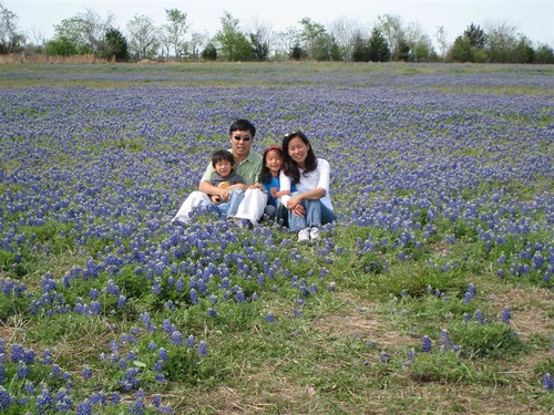
The state flower of Texas is bluebonnets. And they are in season!! We saw fields and patches of these on our trip out to… the Blue Bell Creamery! It was spring break week so we didn’t realize that there would be a LOT of people!

The moms chatted.

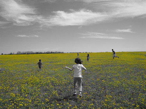
This is the line accentuated with green color.

Max seemed like he was born & ready for Navy!


And they had their own moonwalks!! But it was too hot to jump in them!!


And the family picture… wish I could’ve taken pictures of the inside… but 1) it wasn’t permitted and 2) there wouldn’t have been anything inside worth shooting. The entire tour was a rather disappointing half hour or so. I was hoping for more up-close-and-personal but oh well. It wasn’t a total loss because …
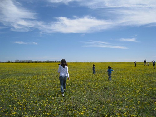


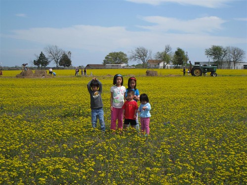
These yellow flowers were EVERYWHERE!! About 4 miles south of Brenham on 290 was this vast HUGE field of yellow flowers!!! They were wild!!
And now onto the … bluebonnets!


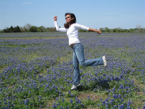
And let us end with this shot of Soojin “trying” to jump into the air! 😀